A comparative study of risk assessment methods for chronic atrophic gastritis combined of spleen deficiency and blood stasis
-
摘要: 目的 分析慢性萎缩性胃炎(chronic atrophic gastritis,CAG)不同风险评估方法的一致性,探索脾虚证、血瘀证量化积分与风险分层的相关性。方法 收集患者的一般信息、症状、舌脉、胃镜及病理资料。根据症状及舌脉进行传统脾虚证、血瘀证诊断及量化评分,根据内镜表现进行脾虚证、血瘀证局部黏膜辨证诊断。采用木村-竹本(K-T)分类、慢性胃炎OLGA(operative link for gastritis assessment)/OLGIM(operative link on gastric intestinal metaplasia assessment)分级对CAG进行风险分层。采用χ2、Kappa一致性检验等分析K-T分类、OLGA/OLGIM评估结果的一致性,考察脾虚证、血瘀证传统辨证与胃镜下黏膜辨证的相关性,分析其与不同风险评估方法的相关性。结果 CAG患者脾虚证、血瘀证积分与萎缩/肠化生范围及程度具有相关性;脾虚证、血瘀证总积分(传统辨证+黏膜辨证)与CAG风险具有相关性,血瘀证量化积分更有助于筛选高风险患者;K-T分类与OLGA/OLGIM分期识别高风险患者结果一致程度中等。结论 木村-竹本分类与OLGA/OLGIM分级分期系统评估结果一致性中等,传统辨证与胃镜局部黏膜辨证结合有助于提高辨证的准确性和全面性,结合脾虚证、血瘀证可进行病证结合风险评估。Abstract: Objective This study aims to analyze the consistency of different risk assessment methods for chronic atrophic gastritis(CAG), and to explore the correlation between the quantitative score of spleen deficiency or blood stasis evidence and the risk of cancer.Methods This study collects patients' general information, endoscopic and pathological data, symptoms, tongue, and pulse diagnosis. The quantized integration of spleen deficiency and blood stasis syndrome is established on the basis of the symptoms, tongue and pulse diagnosis. Diagnosis of endoscopic microdifferentiation of discrimination is based on the endoscopic presentation of spleen deficiency evidence and blood stasis evidence. Risk stratification for CAG was performed with the Kimura-Takemoto (K-T) classification, and the OLGA/OLGIM classification for chronic gastritis.Results There is a correlation between the degree and coverage of spleen deficiency and blood stasis in patients with CAG and the extent of atrophy evaluated by the K-T classification, as well as the severity of histopathological features such as the extent of gastric glandular atrophy (EGA) and intestinal metaplasia (IM). The total score of spleen deficiency and blood stasis evidence (traditional differentiation + endoscopic microdifferentiation of discrimination) correlates with the risk of CAG. Compared with the quantified scoring of spleen deficiency, quantified scoring of blood stasis is more helpful in screening high-risk CAG patients. The agreement between K-T classification and OLGA/OLGIM in identifying high-risk CAG patients by stages is moderate.Conclusion The agreement between K-T classification and OLGA/OLGIM in identifying high-risk CAG patients by stages is moderate. The combination of traditional differentiation and endoscopic microdifferentiation of discrimination helps to improve the accuracy and comprehensiveness of identification. The combination of the evidence of spleen deficiency and blood stasis allows for a risk assessment followed the integration of disease and syndrome.
-

-
表 1 脾虚证、血瘀证胃镜表现
脾虚证胃镜表现 分类 频数(%) 血瘀证胃镜表现 分类 频数(%) 黏膜变薄,苍白或灰白 无 20(32.26) 黏膜下出血点或出血斑 无 22(35.48) 有 42(67.74) 局部 19(30.65) 黏膜下血管清晰可见 无 35(56.45) 多部位 21(33.87) 血管部分透见 16(25.81) 弥漫 0 血管连续均匀 8(12.90) 黏膜暗红色,弥漫性血斑 无 58(93.55) 血管达表层 3(4.84) 有 4(6.45) 黏膜水肿 无 6(9.68) 血管网清晰,色紫暗,树枝样显露 无 45(72.58) 有 56(90.32) 有 17(27.42) 黏膜花斑样改变 无 0 黏膜粗糙不平,呈颗粒样或结节样隆起 无 0 有 62(100.00) 细颗粒/单发结节 23(37.10) 皱襞变细或消失 无 43(69.35) 中等颗粒/多发结节 31(50.00) 有 19(30.65) 粗大颗粒/弥漫结节 8(12.90) 黏液稀薄、清亮 无 30(48.39) 黏膜局限性隆起,顶部凹陷处色泽暗红 无 52(83.87) 有 32(51.61) 单发 6(9.68) 蠕动减弱 无 38(61.29) 多发局部 4(6.45) 有 24(38.71) 多发广泛 0 黏膜弹性减弱 无 41(66.13) 黏液灰白或褐色 无 47(75.81) 有 21(33.87) 有 15(24.19) 表 2 脾虚证、血瘀证症状积分与胃镜表现的关系
X±S 辨证 胃镜表现 无 有 t P 脾虚证 黏膜变薄,苍白或灰白 8.95±5.13 10.36±4.68 -1.073 0.288 黏膜下血管清晰可见 9.77±4.80 10.07±4.96 -0.243 0.809 黏膜水肿 7.67±5.32 10.14±4.77 -1.197 0.236 皱襞变细或消失 9.56±4.73 10.68±5.10 -0.844 0.402 黏液清亮、稀薄 9.57±4.97 10.22±4.76 -0.528 0.600 胃壁蠕动减弱 9.53±5.34 10.50±3.93 -0.770 0.444 黏膜弹性减弱 9.27±4.74 11.14±4.88 -1.458 0.150 血瘀证 黏膜局限性隆起,顶部凹陷处色泽暗红 3.10±2.10 3.70±1.25 -1.229 0.233 黏膜下出血斑或出血点 2.64±1.73 3.50±2.08 -1.658 0.103 黏膜暗红色,弥漫性充血 3.02±1.91 5.75±1.26 -2.801 0.007 血管网清晰,色紫暗,树枝样显露 2.93±1.85 3.88±2.23 -1.701 0.094 黏液灰白或褐色 3.38±1.87 2.60±2.29 1.335 0.187 表 3 萎缩、肠化生程度及部位
例 病变 部位 例数 无 轻度 中度 重度 萎缩 胃窦 60 11 23 22 4 胃角 58 23 15 13 7 胃体 62 28 13 14 7 窦体均有 30 2 4 9 3 肠化生 胃窦 60 8 21 26 5 胃角 58 20 16 14 8 胃体 62 26 15 16 5 窦体均有 32 0 5 11 2 表 4 OLGA与OLGIM
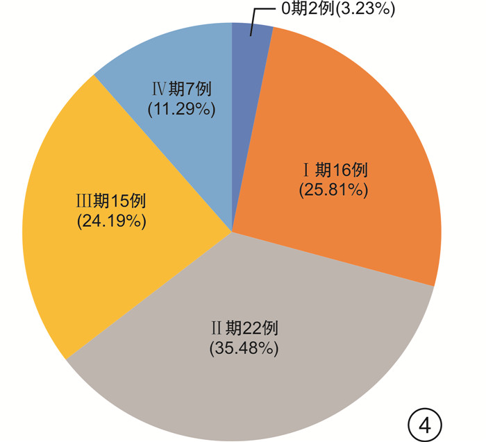
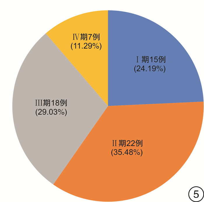
例 评估方法 OLGA 分期 0期 Ⅰ期 Ⅱ期 Ⅲ期 Ⅳ期 风险 低风险 高风险 OLGIM Ⅰ期 0 14 0 1 0 低风险 36 1 Ⅱ期 0 2 20 0 0 高风险 4 21 Ⅲ期 2 0 1 14 1 Ⅳ期 0 0 1 0 6 表 5 K-T分类与OLGA/OLGIM
例 评估方法 OLGA OLGIM 低风险 高风险 低风险 高风险 K-T分类 低风险 29 3 28 4 高风险 11 19 9 21 总计 40 22 37 25 表 6 脾虚证、脾虚血瘀证与CAG风险的相关性
例(%) 评估方法 风险 脾虚证 脾虚血瘀证 χ2 P OLGA 低风险 9(69.23) 27(60.00) 0.365 0.546 高风险 4(30.77) 18(40.00) OLGIM 低风险 7(53.85) 26(57.78) 0.064 0.801 高风险 6(46.15) 19(42.22) K-T分类 低风险 8(61.54) 21(46.67) 0.892 0.345 高风险 5(38.46) 24(53.33) 表 7 脾虚证、血瘀证积分与CAG风险的相关性
X±S 评估方法 辨证积分 低风险 高风险 t P K-T分类 例数 32 30 脾虚证 9.72±4.78 10.10±4.96 -0.308 0.759 血瘀证 2.72±1.78 3.70±2.10 -1.986 0.052 脾虚黏膜辨证 4.16±1.83 5.47±2.00 -2.695 0.009 血瘀黏膜辨证 2.69±1.57 4.47±1.46 -4.611 < 0.001 OLGA 例数 40 22 脾虚证 9.80±5.35 10.09±3.83 -0.225 0.823 血瘀证 2.92±1.79 3.68±2.28 -1.445 0.154 脾虚黏膜辨证 4.17±1.88 5.91±1.77 -3.547 0.001 血瘀黏膜辨证 2.70±1.32 5.09±1.34 -6.77 < 0.001 OLGIM 例数 37 25 脾虚证 9.95±5.54 9.84±3.66 0.045 0.933 血瘀证 3.00±1.68 3.48±2.38 -1.087 0.389 脾虚黏膜辨证 4.19±2.03 5.68±1.65 -3.055 0.003 血瘀黏膜辨证 2.59±1.30 4.96±1.34 -6.944 < 0.001 表 8 脾虚证、血瘀证总积分与CAG风险的相关性
X±S 评估方法 辨证总积分 低风险 高风险 t P OLGA 例数 40 22 脾虚证 13.97±6.18 16.00±3.73 -1.400 0.167 血瘀证 5.63±2.43 8.77±2.81 -4.620 < 0.001 脾虚血瘀证 19.60±6.81 24.77±5.14 -3.106 0.003 OLGIM 例数 37 25 脾虚证 14.14±6.46 15.52±3.60 -1.080 0.285 血瘀证 5.59±2.43 8.44±2.90 -4.179 < 0.001 脾虚血瘀证 19.73±7.10 23.96±5.24 -2.544 0.014 K-T分类 例数 32 30 脾虚证 13.88±5.48 15.57±5.46 -1.216 0.229 血瘀证 5.41±2.45 8.17±2.83 -4.116 < 0.001 脾虚血瘀证 19.28±6.27 23.73±6.48 -2.750 0.008 -
[1] Ajani JA, Bentrem DJ, Besh S, et al. Gastric cancer, version 2.2013: featured updates to the NCCN Guidelines[J]. J Natl Compr Canc Netw, 2013, 11(5): 531-546. doi: 10.6004/jnccn.2013.0070
[2] 陆建邦, 孙喜斌. 解读《中国癌症预防与控制规划纲要(2004—2010)》[J]. 中国肿瘤, 2004, 13(6): 342-344. doi: 10.3969/j.issn.1004-0242.2004.06.003
[3] 国家消化系疾病临床医学研究中心(上海), 国家消化道早癌防治中心联盟, 中华医学会消化病学分会幽门螺杆菌学组, 等. 中国胃黏膜癌前状态和癌前病变的处理策略专家共识(2020年)[J]. 中华消化杂志, 2020, 40(11): 731-741. doi: 10.3760/cma.j.cn311367-20200915-00554
[4] Sugano K, Tack J, Kuipers EJ, et al. Kyoto global consensus report on Helicobacter pylori gastritis[J]. Gut, 2015, 64(9): 1353-1367. doi: 10.1136/gutjnl-2015-309252
[5] 房静远, 杜奕奇, 刘文忠, 等. 中国慢性胃炎共识意见(2017年, 上海)[J]. 胃肠病学, 2017, 22(11): 670-687. doi: 10.3969/j.issn.1008-7125.2017.11.007
[6] 张声生, 胡玲, 李茹柳. 脾虚证中医诊疗专家共识意见(2017)[J]. 中医杂志, 2017, 58(17): 1525-1530.
[7] 杜金行, 史载祥. 血瘀证中西医结合诊疗共识[J]. 中国中西医结合杂志, 2011, 31(6): 839-844.
[8] 中国中西医结合学会活血化瘀专业委员会. 实用血瘀证诊断标准[J]. 中国中西医结合杂志, 2016, 36(10): 1163. doi: 10.7661/CJIM.2016.10.1163
[9] 中华医学会消化内镜学分会. 慢性胃炎的内镜分型分级标准及治疗的试行意见[J]. 中华消化内镜杂志, 2004, 21(2): 4-5.
[10] 张声生, 唐旭东, 黄穗平, 等. 慢性胃炎中医诊疗专家共识意见(2017)[J]. 中华中医药杂志, 2017, 32(7): 3060-3064.
[11] 陈家旭, 邹小娟. 中医诊断学[M]. 2版. 北京: 人民卫生出版社. 2014: 38-45.
[12] Vannella L, Lahner E, Osborn J, et al. Risk factors for progression to gastric neoplastic lesions in patients with atrophic gastritis[J]. Aliment Pharmacol Ther, 2010, 31(9): 1042-1050.
[13] González CA, Pardo ML, Liso JM, et al. Gastric cancer occurrence in preneoplastic lesions: a long-term follow-up in a high-risk area in Spain[J]. Int J Cancer, 2010, 127(11): 2654-2660. doi: 10.1002/ijc.25273
[14] 刘赓, 丁洋, 张声生. 张声生从"虚"、"毒"、"瘀"论治慢性萎缩性胃炎[J]. 中国中医基础医学杂志, 2012, 18(10): 1098-1099.
[15] 蔡淦. 慢性萎缩性胃炎及其癌前病变的中医治疗[J]. 江苏中医药, 2007, 39(8): 1.
[16] 王爱云, 单兆伟. 慢性萎缩性胃炎从血瘀论治[J]. 中国中西医结合脾胃杂志, 2000, 8(5): 290-291.
[17] 刘赓, 唐旭东. 唐旭东辨证治疗慢性萎缩性胃炎经验体会[J]. 辽宁中医杂志, 2009, 36(5): 734-736.
[18] 吴丹. 慢性萎缩性胃炎胃镜象与证素特点的相关性研究[D]. 福州: 福建中医药大学, 2018.
[19] 董丽霞. 慢性胃炎胃镜下微观表现与中医宏观辨证的相关性研究[D]. 济南: 山东中医药大学, 2016.
[20] 许话. 慢性萎缩性胃炎中医证型分布与胃镜象、病理象相关性的研究[D]. 北京: 北京中医药大学, 2014.
[21] 黄雅慧, 郭菊清, 刘越洋, 等. 慢性萎缩性胃炎胃黏膜癌前病变病理变化与中医证型及Hp的相关性研究[J]. 中华中医药学刊, 2014, 32(6): 1381-1383.
[22] 李莉, 朱蕾蕾, 孙祝美, 等. 慢性萎缩性胃炎中医证型分布及幽门螺杆菌感染、胃黏膜病理变化情况分析[J]. 上海中医药杂志, 2019, 52(6): 20-23.
[23] 黄远程, 潘静琳, 黄超原, 等. 慢性萎缩性胃炎癌前病变证型、证素演变规律文献研究[J]. 中医杂志, 2019, 60(20): 1778-1783.
[24] 杨洋, 瞿先侯, 杨敏, 等. 慢性萎缩性胃炎患者中医证候分型与癌变风险的相关性[J]. 中医杂志, 2020, 61(4): 319-324.
[25] 李军祥, 陈誩, 吕宾, 等. 慢性萎缩性胃炎中西医结合诊疗共识意见(2017年)[J]. 中国中西医结合消化杂志, 2018, 26(2): 121-131.
[26] 张贺军, 金珠, 崔荣丽, 等. OLGA分期、分级评估系统在胃镜活检组织病理学评价中的应用[J]. 中华消化内镜杂志, 2014, 31(3): 121-125.
[27] Cotruta B, Gheorghe C, Iacob R, et al. The orientation of gastric biopsy samples improves the inter-observer agreement of the OLGA staging system[J]. J Gastrointestin Liver Dis, 2017, 26(4): 351-356.
[28] 陈璇, 瞿旻晔, 史肖华, 等. 木村-竹本分类法在胃癌筛查中的应用[J]. 现代医学, 2020, 48(9): 1238-1241.
[29] 李兆申, 王贵齐, 张澍田, 等. 中国早期胃癌筛查流程专家共识意见(草案)(2017年, 上海)[J]. 胃肠病学, 2018, 23(2): 92-97. https://xuewen.cnki.net/CCND-JFRB202211070012.html
-


 下载:
下载:
