Clinic correlation and prognostic value of P4HB and GRP78 expression in gastric cancer
-
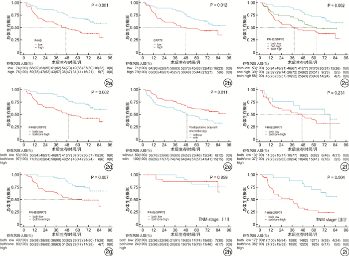
摘要: 目的 探讨脯氨酰4-羟化酶β多肽(P4HB)和葡萄糖调节蛋白78(GRP78)表达与胃癌临床特征的相关性及对患者预后的预测价值。方法 采用免疫组织化学法分别评估150例胃癌组织样本中P4HB和GRP78蛋白的表达,分析蛋白表达与胃癌临床病理学特征的关联。采用Kaplan-Meier分析比较总生存期(OS)的生存曲线。用单变量和多变量Cox回归模型分析影响OS的潜在预后因素。基于多变量Cox回归模型构建预后列线图,并与TNM分期比较其临床价值。结果 P4HB的表达与患者年龄、Bormann分型、肿瘤浸润深度、淋巴结转移、术后辅助化疗相关,GRP78的表达与肿瘤浸润深度和淋巴结转移相关;两者的表达呈正相关。Kaplan-Meier分析表明,P4HB和GRP78的高单一表达或共表达预示较短的OS,两者的高共表达在术后辅助化疗组,特别是晚期组中预后不良。多因素Cox回归分析确定癌组织分化程度、TNM分期、术后辅助化疗、P4HB与GRP78共表达是OS的独立预后因素。在受试者工作特征曲线和决策曲线分析中,列线图在辨别能力和临床实用性方面优于TNM分期。结论 P4HB与GRP78的表达呈正相关,其共表达是OS的独立预后因素,可作为胃癌患者特别是晚期术后辅助化疗患者OS的预测生物标志物。预后列线图模型可能具有较高的临床应用价值。
-
关键词:
- 脯氨酰4-羟化酶β多肽 /
- 葡萄糖调节蛋白78 /
- 胃癌 /
- 术后辅助化疗 /
- 列线图
Abstract: Objective To explore the correlation and prognostic value of prolyl 4-hydroxylase beta polypeptide(P4HB) and glucose-regulated protein 78(GRP78) expressions combined with clinical features in gastric cancer.Methods One hundred and fifty gastric cancer tissue samples were evaluated P4HB and GRP78 protein expressions by immunohistochemistry separately. Association of the expressions with clinicopathological features was analyzed. Kaplan-Meier analyses were taken to compare survival curves of overall survival(OS). Univariate and multivariate Cox regression models were used to analyze potential prognostic factors of OS. Basing on the multivariate Cox regression model, a prognostic nomogram was constructed, clinical usefulness of which was compared to TNM stage.Results The expressions of P4HB were correlated with age, Bormann type, depth of tumor invasion, lymph node metastasis, postoperative adjuvant chemotherapy, while the expressions of GRP78 were correlated with the depth of tumor invasion and lymph node metastasis. There was a positive correlation between their expressions. According to Kaplan-Meier analyses, high single expression or co-expression of P4HB and GRP78 indicated a shorter OS, high co-expression represented an unfavorable prognosis in the group with postoperative adjuvant chemotherapy, especially in the advanced stage. Multivariate Cox regression analysis identified differentiation, TNM stage, postoperative adjuvant chemotherapy, P4HB and GRP78 co-expression were independent prognostic factors for OS. The nomogram was better than TNM stage in discrimination ability and clinical usefulness shown in the receiver operating characteristic curves and decision curve analysis curves.Conclusion P4HB was positively correlated with GRP78 expression, co-expression of them was an independent prognostic factor, and could serve as a predictive biomarker for gastric cancer patients of OS, especially for advanced stage patients with postoperative adjuvant chemotherapy. The prognostic nomogram model may have a high clinical application value. -

-
表 1 胃癌患者P4HB、GRP78表达与临床病理特征的关系
例 临床病理特征 P4HB表达 GRP78表达 低 高 χ2 P 低 高 χ2 P 总体 74 76 71 79 年龄/岁 < 65 48 35 5.369 0.021 44 39 2.404 0.121 ≥65 26 41 27 40 性别 男 49 54 0.408 0.523 49 54 0.008 0.931 女 25 22 22 25 Bormann分型 Ⅰ~Ⅱ 32 20 4.743 0.029 25 27 0.018 0.894 Ⅲ~Ⅳ 42 56 46 52 肿瘤大小/cm < 5 43 40 0.455 0.500 41 42 0.318 0.573 ≥5 31 36 30 37 Lauren分型 肠型 17 14 12 19 弥漫型 40 31 5.489 0.064 38 33 2.263 0.323 混合型 17 31 21 27 分化程度 高、中分化 31 29 0.218 0.641 29 31 0.040 0.841 低分化 43 47 42 48 病理类型 腺癌 66 73 2.599 0.107 63 76 3.071 0.080 黏液、印戒细胞癌 8 3 8 3 肿瘤浸润深度 T1~T3 61 43 11.786 0.001 55 49 4.192 0.041 T4 13 33 16 30 淋巴结转移 阴性 32 19 5.561 0.018 30 21 4.092 0.043 阳性 42 57 41 58 TNM分期 Ⅰ~Ⅱ 41 31 3.209 0.073 37 35 0.914 0.339 Ⅲ~Ⅳ 33 45 34 44 血管浸润 阴性 53 53 0.064 0.800 48 58 0.609 0.435 阳性 21 23 23 21 周围神经浸润 阴性 56 59 0.080 0.777 54 61 0.028 0.867 阳性 18 17 17 18 术后辅助化疗 无 17 33 7.055 0.008 22 28 0.334 0.563 有 57 43 59 51 GRP78表达 低 53 18 34.562 < 0.001 高 21 58 表 2 胃癌患者OS预后特征的单因素和多因素Cox回归分析
相关因素 单因素分析 多因素分析 HR 95%CI P HR 95%CI P 年龄/岁(≥65 vs. < 65) 1.246 0.791~1.963 0.343 性别(女vs.男) 1.081 0.665~1.756 0.754 Bormann分型(Ⅲ~Ⅳ vs.Ⅰ~Ⅱ) 2.142 1.271~3.609 0.004 肿瘤大小/cm(≥5 vs. < 5) 1.662 1.056~2.616 0.028 Lauren分型 0.214 弥漫型vs.肠型 1.818 0.932~3.545 0.079 混合型vs.肠型 1.613 0.790~3.293 0.190 分化程度(低分化vs.高、中分化) 2.111 1.274~3.497 0.004 1.846 1.099~3.101 0.020 病理类型[黏液(印戒)细胞癌vs.腺癌] 0.592 0.216~1.622 0.308 肿瘤浸润深度(T4 vs.T1~T3) 2.536 1.601~4.019 < 0.001 淋巴结转移(阳性vs.阴性) 3.824 2.060~7.101 < 0.001 TNM分期(Ⅲ~Ⅳ vs.Ⅰ~Ⅱ) 3.200 1.942~5.275 < 0.001 3.124 1.870~5.217 < 0.001 血管浸润(阳性vs.阴性) 1.385 0.861~2.229 0.180 周围神经浸润(阳性vs.阴性) 1.279 0.766~2.135 0.346 术后辅助化疗(有vs.无) 0.556 0.351~0.879 0.012 0.507 0.317~0.810 0.005 P4HB表达(高vs.低) 2.266 1.414~3.630 0.001 GRP78表达(高vs.低) 1.799 1.130~2.862 0.013 P4HB和GRP78共表达(两者/一者高vs.两者低) 2.260 1.338~3.817 0.002 2.304 1.355~3.915 0.002 -
[1] Sung H, Ferlay J, Siegel RL, et al. Global Cancer Statistics 2020: GLOBOCAN Estimates of Incidence and Mortality Worldwide for 36 Cancers in 185 Countries[J]. CA Cancer J Clin, 2021, 71(3): 209-249. doi: 10.3322/caac.21660
[2] 秦虹, 宋金霞, 底玮, 等. 胃癌组织中的DAB2IP、Snail表达及其与临床病理特征和预后的关系[J]. 中国中西医结合消化杂志, 2021, 29(7): 411-415. doi: 10.3969/j.issn.1671-038X.2021.06.06
[3] Jiang Y, Zhang Q, Hu Y, et al. ImmunoScore Signature: A Prognostic and Predictive Tool in Gastric Cancer[J]. Ann Surg, 2018, 267(3): 504-513. doi: 10.1097/SLA.0000000000002116
[4] Joshi SS, Badgwell BD. Current treatment and recent progress in gastric cancer[J]. CA Cancer J Clin, 2021, 71(3): 264-279. doi: 10.3322/caac.21657
[5] Tameire F, Verginadis Ⅱ, Koumenis C, et al. Cell intrinsic and extrinsic activators of the unfolded protein response in cancer: Mechanisms and targets for therapy[J]. Semin Cancer Biol, 2015, 33: 3-15. doi: 10.1016/j.semcancer.2015.04.002
[6] Kranz P, Neumann F, Wolf A, et al. PDI is an essential redox-sensitive activator of PERK during the unfolded protein response(UPR)[J]. Cell Death Dis, 2017, 8(8): e2986. doi: 10.1038/cddis.2017.369
[7] Wang M, Kaufman RJ. The impact of the endoplasmic reticulum protein-folding environment on cancer development[J]. Nat Rev Cancer, 2014, 14(9): 581-597. doi: 10.1038/nrc3800
[8] Zou H, Wen C, Peng Z, et al. P4HB and PDIA3 are associated with tumor progression and therapeutic outcome of diffuse gliomas[J]. Oncol Rep, 2018, 39(2): 501-510.
[9] Shi R, Gao S, Zhang J, et al. Collagen prolyl 4-hydroxylases modify tumor progression[J]. Acta Biochim Biophys Sin(Shanghai), 2021, 53(7): 805-814.
[10] Kong Y, Chen H, Chen M, et al. Abnormal ECA-Binding Membrane Glycans and Galactosylated CAT and P4HB in Lesion Tissues as Potential Biomarkers for Hepatocellular Carcinoma Diagnosis[J]. Front Oncol, 2022, 12: 855952. doi: 10.3389/fonc.2022.855952
[11] Zhang J, Wu Y, Lin YH, et al. Prognostic value of hypoxia-inducible factor-1 alpha and prolyl 4-hydroxylase beta polypeptide overexpression in gastric cancer[J]. World J Gastroenterol, 2018, 24(22): 2381-2391. doi: 10.3748/wjg.v24.i22.2381
[12] Kwon D, Koh J, Kim S, et al. Overexpression of endoplasmic reticulum stress-related proteins, XBP1 s and GRP78, predicts poor prognosis in pulmonary adenocarcinoma[J]. Lung Cancer, 2018, 122: 131-137. doi: 10.1016/j.lungcan.2018.06.005
[13] Gifford JB, Hill R. GRP78 Influences Chemoresistance and Prognosis in Cancer[J]. Curr Drug Targets, 2018, 19(6): 701-708. doi: 10.2174/1389450118666170615100918
[14] Nagelkerke A, Bussink J, Sweep FC, et al. The unfolded protein response as a target for cancer therapy[J]. Biochim Biophys Acta, 2014, 1846(2): 277-284.
[15] Gonzalez-Gronow M, Pizzo SV. Physiological Roles of the Autoantibodies to the 78-Kilodalton Glucose-Regulated Protein(GRP78) in Cancer and Autoimmune Diseases[J]. Biomedicines, 2022, 10(6): 1222. doi: 10.3390/biomedicines10061222
[16] Xia W, Zhuang J, Wang G, et al. P4HB promotes HCC tumorigenesis through downregulation of GRP78 and subsequent upregulation of epithelial-to-mesenchymal transition[J]. Oncotarget, 2017, 8(5): 8512-8521. doi: 10.18632/oncotarget.14337
[17] Li L, Cui Y, Ji JF, et al. Clinical Correlation Between WISP2 and beta-Catenin in Gastric Cancer[J]. Anticancer Res, 2017, 37(8): 4469-4473.
[18] Mohi-Ud-Din R, Mir RH, Wani TU, et al. The Regulation of Endoplasmic Reticulum Stress in Cancer: Special Focuses on Luteolin Patents[J]. Molecules, 2022, 27(8): 2471. doi: 10.3390/molecules27082471
[19] Lee E, Lee DH. Emerging roles of protein disulfide isomerase in cancer[J]. BMB Rep, 2017, 50(8): 401-410. doi: 10.5483/BMBRep.2017.50.8.107
[20] Gwark S, Ahn HS, Yeom J, et al. Plasma Proteome Signature to Predict the Outcome of Breast Cancer Patients Receiving Neoadjuvant Chemotherapy[J]. Cancers(Basel), 2021, 13(24): 6267.
[21] Casas C. GRP78 at the Centre of the Stage in Cancer and Neuroprotection[J]. Front Neurosci, 2017, 11: 177.
[22] Ibrahim IM, Abdelmalek DH, Elfiky AA. GRP78: A cell's response to stress[J]. Life Sci, 2019, 226: 156-163. doi: 10.1016/j.lfs.2019.04.022
[23] Dai YJ, Qiu YB, Jiang R, et al. Concomitant high expression of ERalpha36, GRP78 and GRP94 is associated with aggressive papillary thyroid cancer behavior[J]. Cell Oncol(Dordr), 2018, 41(3): 269-282. doi: 10.1007/s13402-017-0368-y
[24] Liu M, Yang J, Lv W, et al. Down-regulating GRP78 reverses pirarubicin resistance of triple negative breast cancer by miR-495-3p mimics and involves the p-AKT/mTOR pathway[J]. Biosci Rep, 2022, 42(1): 20210245. doi: 10.1042/BSR20210245
[25] Foth M, Mcmahon M. Autophagy Inhibition in BRAF-Driven Cancers[J]. Cancers(Basel), 2021, 13(14): 3498.
[26] Mayer M, Kies U, Kammermeier R, et al. BiP and PDI cooperate in the oxidative folding of antibodies in vitro[J]. J Biol Chem, 2000, 275(38): 29421-29425. doi: 10.1074/jbc.M002655200
[27] Kim C, Kim B. Anti-Cancer Natural Products and Their Bioactive Compounds Inducing ER Stress-Mediated Apoptosis: A Review[J]. Nutrients, 2018, 10(8): 1021. doi: 10.3390/nu10081021
[28] Wang Y, Wang K, Jin Y, et al. Endoplasmic reticulum proteostasis control and gastric cancer[J]. Cancer Lett, 2019, 449: 263-271.
[29] Khaled J, Kopsida M, Lennernas H, et al. Drug Resistance and Endoplasmic Reticulum Stress in Hepatocellular Carcinoma[J]. Cells, 2022, 11(4): 632. doi: 10.3390/cells11040632
[30] Shi Z, Yu X, Yuan M, et al. Activation of the PERK-ATF4 pathway promotes chemo-resistance in colon cancer cells[J]. Sci Rep, 2019, 9(1): 3210. doi: 10.1038/s41598-019-39547-x
-


 下载:
下载:


