
Establishment and evaluation of a combination rat model of Barrett esophagus combined with reflux disease
-
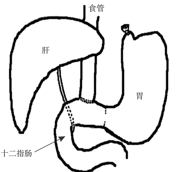
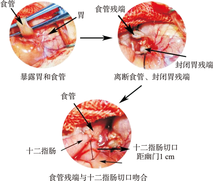
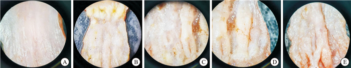
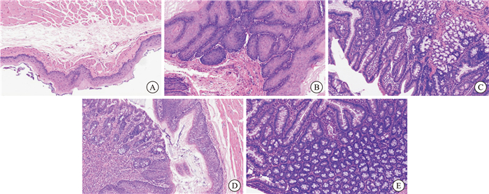
摘要: 目的 建立一种Barrett食管(Barrett esophagus,BE)酸碱混合反流的大鼠模型。 方法 将50只Wistar雄性大鼠随机分为对照组(10只)和模型组(40只)。将模型组随机分为2组(模型甲、乙组),每组20只,均采用食管-胃-十二指肠吻合术进行造模。模型甲组手术操作过程中先进行食管-十二指肠吻合术,吻合1/2圈后进行十二指肠纵行切开,再吻合其余部分;模型乙组手术操作过程中先将十二指肠纵行切开,再行食管-十二指肠吻合。观察模型甲、乙组术后1周大鼠体重及存活率。对存活率较高的模型组,将该组存活大鼠随机分为2组(模型丙、丁组),在术后第4周,模型丙组不予任何处理,模型丁组予右旋糖酐铁(4 mg/kg/周)腹腔注射;分别饲养22周。考虑样本量及动物伦理情况,对模型丙、丁组再另外各追加10只大鼠扩充样本量,随机分组按照上述造模方式进行造模。在饲养期间对大鼠的精神行为、体重、进食量、饮水量及抓力等方面进行测量,从多维度进行综合评价。分别于术后16周、22周处死部分大鼠,对大鼠食管组织进行肉眼观察及病理组织学观察,判断建模是否成功,确定最佳建模方法及建立周期。 结果 模型甲、乙组大鼠术后生活质量良好,但体重均较对照组明显下降(P < 0.01),且模型乙组死亡率(20%)较模型甲组低(40%)。将模型甲组随机分为2组(模型丙、丁组),每组8只,术后16周及22周模型丙组与丁组之间大鼠体重、进食量、饮水量及抓力等方面均较对照组下降(P < 0.05);模型丙与丁组间,术后22周与16周组内上述指标均差异无统计学意义(P>0.05)。模型丙、丁组大鼠食管肉眼均可见到不同程度的黏膜充血、水肿伴斑块状突起,部分可见糜烂灶,且模型丁组术后16周和22周可见到部分黏膜呈橘红色,22周时更为明显。在模型丙组术后22周、模型丁组术后16周、模型丁组术后22周大鼠食管下端病理组织学苏木精-伊红染色均可见到柱状上皮化生,且模型丁组术后22周时最为显著。 结论 食管-胃-十二指肠吻合术联合右旋糖酐铁腹腔注射方法进行BE造模,术后22周时成模率最高,术后并发症及死亡率相对较小。Abstract: Objective To explore and establish a rat model of acid and alkaline mixed reflux in Barrett esophagus(BE) through multi-faceted evaluation. Methods Fifty male Wistar rats were randomly divided into a normal control group(10 rats) and a model group(40 rats). The model group was further randomly divided into two subgroups(20 rats each). Both subgroups underwent esophago-gastro-duodenostomy(EGDA) for modeling. In subgroup A, esophago-duodenostomy was performed first, followed by a longitudinal incision of the duodenum and completion of the anastomosis. In subgroup B, the duodenum was incised longitudinally first, followed by esophago-duodenostomy. The body weight and survival rate of the rats in both subgroups were observed one week after the operation. For the subgroup with a higher survival rate, the surviving rats were randomly divided into two groups(subgroup C and D). At the fourth week after the operation, subgroup C received no treatment, while subgroup D was given intraperitoneal injection of iron dextran(4 mg/kg/week). The rats were raised for 22 weeks. Considering the sample size and animal ethics, an additional 10 rats were added to each of the model groups C and D to increase the sample size. They were randomly grouped and modeled according to the above-mentioned modeling methods. During the raising period, the mental behavior, body weight, food intake, water intake, and grip strength of the rats were measured for comprehensive evaluation. At 16 and 22 weeks after the operation, some rats were sacrificed, and the esophageal tissues were observed macroscopically and histopathologically to determine the success of modeling and the optimal modeling method and cycle. Results The postoperative quality of life of rats in subgroups A and B was good, but their body weight was significantly lower than that of the control group(P < 0.01), and the mortality rate of subgroup B(20%) was lower than that of subgroup A(40%). Subgroup A was randomly divided into two groups(subgroup C and D), with 8 rats in each group. At 16 and 22 weeks after the operation, the body weight, food intake, water intake, and grip strength of rats in subgroups C and D were all lower than those of the control group(P < 0.05). There was no significant statistical difference in the above aspects between subgroups C and D at 22 and 16 weeks after the operation(P>0.05). Macroscopic observation of the esophagus in subgroups C and D showed varying degrees of mucosal congestion, edema, and plaque-like protrusions, and some had erosions. At 16 and 22 weeks after the operation in subgroup D, some mucosa was orange-red, which was more obvious at 22 weeks. Histopathological observation of the esophagus in subgroups C at 22 weeks after the operation, subgroup D at 16 weeks after the operation, and subgroup D at 22 weeks after the operation all showed goblet cell infiltration, and it was most significant in subgroup D at 22 weeks after the operation. Conclusion The method of esophago-gastro-duodenostomy combined with intraperitoneal injection of iron dextran for BE modeling has the highest success rate at 22 weeks after the operation, with relatively fewer postoperative complications and mortality.
-
Key words:
- Barrett esophagus /
- acid-base mixed reflux /
- model /
- iron
-

-
表 1 各组大鼠术后体重变化
g,X±S 组别 例数 术后16周 术后22周 对照组 5 327.82±8.73 351.22±9.03 模型丙组 8 242.83±10.011) 272.35±5.041) 模型丁组 7 202.34±11.121)3) 254.32±4.711)2) 与对照组比较,1)P < 0.01;与模型丙组比较,2)P < 0.05,3)P < 0.01。  下载: 导出CSV
表 2 各组大鼠术后进食量变化
g,X±S 组别 例数 术后16周 术后22周 对照组 5 35.25±0.99 36.30±0.59 模型丙组 8 30.05±1.091) 31.47±1.291) 模型丁组 7 28.07±0.631) 27.14±1.031)2) 与对照组比较,1)P < 0.01;与模型丙组比较,2)P < 0.01。
下载: 导出CSV
表 3 各组大鼠术后饮水量变化
mL,X±S 组别 例数 术后16周 术后22周 对照组 5 36.09±0.95 38.92±1.85 模型丙组 8 31.03±0.691) 29.86±0.601) 模型丁组 7 28.63±0.941)2) 26.34±1.671) 与对照组比较,1)P < 0.01;与模型丙组比较,2)P < 0.05。
下载: 导出CSV
表 4 各组大鼠术后抓力变化
g,X±S 组别 例数 术后16周 术后22周 对照组 5 1 499.12±67.33 1 611.81±123.20 模型丙组 8 1 229.24±45.681) 1 053.10±73.491) 模型丁组 7 1 188.57±40.761) 916.02±79.811) 与对照组比较,1)P < 0.01。
下载: 导出CSV
-
[1] 中华医学会消化病学分会. 2020年中国胃食管反流病专家共识[J]. 中华消化杂志, 2020, 40(10): 649-663. doi: 10.3760/cma.j.cn311367-20200918-00558
[2] Mohy-Ud-Din N, Krill TS, Shah AR, et al. Barrett's esophagus: what do we need to know?[J]. Dis Mon, 2020, 66(1): 100850. doi: 10.1016/j.disamonth.2019.02.003
[3] 朱称心, 蔡林, 张兴龙, 等. 食管癌筛查的研究进展[J]. 中华肿瘤防治杂志, 2024, 31(22): 1399-1405.
[4] Cotton CC, Eluri S, Shaheen NJ. Management of dysplastic barrett's esophagus and early esophageal adenocarcinoma[J]. Gastroenterol Clin North Am, 2022, 51(3): 485-500. doi: 10.1016/j.gtc.2022.06.004
[5] Thorell A, Bednarska O, Jeremiasen M, et al. Barrett's esophagus: Diagnosis, treatment and minimizing of risk[J]. Lakartidningen, 2023, 120: 23076.
[6] 郭政, 唐艳萍, 李明莉, 等. 从反流性食管炎到巴雷特食管: 食管菌群扮演的角色[J]. 中国中西医结合消化杂志, 2023, 31(10): 810-815. doi: 10.3969/j.issn.1671-038X.2023.10.16
[7] Beydoun AS, Stabenau KA, Altman KW, et al. Cancer risk in barrett's esophagus: a clinical review[J]. Int J Mol Sci, 2023, 24(7): 6018. doi: 10.3390/ijms24076018
[8] Äkerström JH, Santoni G, von Euler Chelpin M, et al. Antireflux surgery versus antireflux medication and risk of esophageal adenocarcinoma in patients with barrett's esophagus[J]. Gastroenterology, 2024, 166(1): 132-138. e3. doi: 10.1053/j.gastro.2023.08.050
[9] 胡小海. 巴雷特食管类器官体外模型的构建及ARID1A对巴雷特食管发生的影响[D]. 沈阳: 中国医科大学, 2024.
[10] Maslyonkina KS, Konyukova AK, Alexeeva DY, et al. Barrett's esophagus: The pathomorphological and molecular genetic keystones of neoplastic progression[J]. Cancer Med, 2022, 11(2): 447-478. doi: 10.1002/cam4.4447
[11] 刘磊, 唐艳萍, 弓艳霞, 等. 活血通降方对反流性食管炎大鼠食管黏膜NF-κB/MAPK信号转导通路的影响[J]. 中国中西医结合外科杂志, 2021, 27(4): 555-562. doi: 10.3969/j.issn.1007-6948.2021.04.002
[12] 陈小苏, 王艳, 鲍云, 等. 胃食管反流病大鼠模型探讨[J]. 中华胃食管反流病电子杂志, 2020, 7(4): 228-235.
[13] 刘磊, 郭政, 唐艳萍, 等. 基于网络药理学和分子对接探究活血通降方治疗反流性食管炎的分子机制[J]. 中国中西医结合外科杂志, 2024, 30(5): 722-728.
-
计量
- 文章访问数: 206
- 施引文献: 0