
Diagnostic value of narrow band imaging and magnifying endoscopy in gastric neuroendocrine neoplasms
-
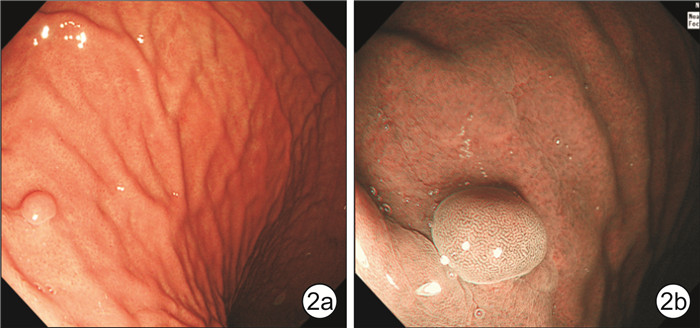
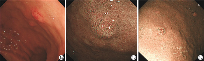
摘要: 目的 分析运用白光内镜(WLI)及放大内镜窄带成像(ME-NBI)诊断胃神经内分泌肿瘤(G-NENs)的特点并探讨其临床诊断价值。方法 选择2017年1月-2021年6月西安市人民医院收治的36例G-NENs患者(观察组)及同期收治的40例胃息肉患者(对照组)临床资料,分析观察组临床病理特点及2组病变ME-NBI特点。结果 WLI结合ME-NBI诊断G-NENs敏感性为80.6%,特异性为90.0%,阳性预测值为87.9%,阴性预测值为83.7%,准确度为85.5%,约登指数为0.71。结论 WLI结合ME-NBI有利于观察G-NENs表面特异性的腺体结构改变及上皮下黑棕色迂曲增粗的螺旋状血管,对于鉴别G-NENs及胃息肉有重要意义,能够针对性地对病灶进行靶向活检,避免漏诊及误诊,并对正确治疗方式选择提供帮助。Abstract: Objective To investigate the values of white light imaging combined with magnifying endoscopy and narrow band imaging(ME-NBI) in the diagnose of the gastric neuroendocrine neoplasms(G-NENs).Methods Clinical data, characteristics of WLI and ME-NBI, pathological and immunehistochemical results of 36 G-NENs patients and 40 gastric polyps patients from January 2017 to June 2021 in our hospital were collected and retrospectively analyzed.Results WLI combined with ME-NBI diagnosis of G-NENs showed that the sensitivity was 80.6%, the specificity was 90.0%, the positive predictive value was 87.9%, the negative predictive value was 83.7%, the accuracy rating was 85.5% and the Yoden index was 0.71.Conclusion WLI combined with ME-NBI has been shown to be more effective for targeting biopsy by discovery specific structural changes and dilated blackish-brown subepithelial vessels with cork-screw capillaries on the surface of G-NENs, has a significant clinical value for diagnosis of G-NEN and differentiation diagnosis of gastric polyps, avoid and reduce the rate of misdiagnose and adapts appropriate treatment strategies.
-

-
表 1 患者基线资料及内镜下特点
类别 观察组(n=36) 对照组(n=40) P 年龄/岁 47.0±3.1 45.0±2.6 0.332 性别(男/女) 16/20 19/21 0.821 胃泌素/(pg·mL-1) 297.2(238.8,408.8) 105.6(59.2,170.9) 0.004 临床症状/例 0.720 腹胀 20 22 腹痛 16 18 WLI/例 色泽发红 30 3 < 0.001 ME-NBI/例 微结构改变 21 0 < 0.001 微血管改变 30 4 < 0.001  下载: 导出CSV
-
[1] 李勇, 王勇飞, 檀碧波, 等. 355例胃肠胰神经内分泌肿瘤的临床病理特征与生存分析[J]. 中华肿瘤杂志, 2020, 42(5): 426-431. doi: 10.3760/cma.j.cn112152-112152-20191011-00663
[2] 苏惠, 李娜, 王海红, 等. 167例胃肠道神经内分泌肿瘤的内镜表现及病理特征回顾性分析[J]. 胃肠病学和肝病学杂志, 2019, 28(4): 405-409. https://www.cnki.com.cn/Article/CJFDTOTAL-WCBX201904011.htm
[3] Chung CS, Tsai CL, Yin Y, et al. Clinical features and outcomes of gastric neuroendocrine tumors after endoscopic diagnosis and treatment: A Digestive Endoscopy Society of Tawian(DEST)[J]. Medicine, 2018, 97(38): e12101. doi: 10.1097/MD.0000000000012101
[4] 邱旭东, 刘猛, 刘青, 等. 903例神经内分泌肿瘤发病部位与病理特征分析[J]. 中华胃肠外科杂志, 2017, 20(9): 993-996. doi: 10.3760/cma.j.issn.1671-0274.2017.09.008
[5] Fan JH, Zhang YQ, Shi SS, et al. A nation-wide retrospective epidemiological study of gastroenteropancreatic neuroendocrine neoplasms in china[J]. Oncotarget, 2017, 8(42): 71699-71708. doi: 10.18632/oncotarget.17599
[6] Fang C, Wang W, Zhang Y, et al. Clinicopathologic characteristics and prognosis of gastroenteropancreatic neuroendocrine neoplasms: a multicenter study in South China[J]. Chin J Cancer, 2017, 36(10): 497-505. https://cancercommun.biomedcentral.com/articles/10.1186/s40880-017-0218-3
[7] WHO classification of Tumors Editorial Board. WHO classification of tumors Digestive system tumors[M], Lyon: IRAC Press, 2019: 14-20.
[8] 中华医学会病理学分会消化疾病学组, 2020年中国胃肠胰神经内分泌肿瘤病理诊断共识专家组. 中国胃肠胰神经内分泌肿瘤病理诊断共识(2020版)[J]. 中华病理学杂志, 2021, 50(1): 14-20.
[9] Nagtegaal ID, Odze RD, Klimstra D, et al. The 2019. WHO classification of tumours of digestive system[J]. Histopathology, 2020, 76(2): 182-188. doi: 10.1111/his.13975
[10] 罗杰, 史艳芬, 谭煌英. 胃神经内分泌肿瘤临床分型与病理[J]. 中华消化杂志, 2019, 39(8): 516-520. doi: 10.3760/cma.j.issn.0254-1432.2019.08.004
[11] 李远良, 罗杰, 谭煌英. 胃神经内分泌肿瘤的分型诊治进展[J]. 临床肿瘤学杂志, 2020, 25(9): 857-861. doi: 10.3969/j.issn.1009-0460.2020.09.014
[12] Lo GC, Kambadakone A. MR imaging of pancreatic neuroendocrine tumors[J]. Magn Reson Imaging Clin N Am, 2018, 26(3): 391-403. doi: 10.1016/j.mric.2018.03.010
[13] 夏盛伟, 余捷, 林细州, 等. 胃神经内分泌肿瘤CT检查影像学特征[J]. 中华消化外科杂志, 2020, 19(9): 995-1000. doi: 10.3760/cma.j.cn115610-20200814-00551
[14] Hofland J, Zandee WT, de Herder WW. Role of biomarker tests for diagnosis of neuroendocrine tumours[J]. Nat Rev Endocrinol, 2018, 14(11): 656-669. doi: 10.1038/s41574-018-0082-5
[15] Monjur A. Gastrointestinal neuroendocrine tumors in 2020[J]. World J Gastrointest Oncol, 2020, 12(8): 791-807. doi: 10.4251/wjgo.v12.i8.791
[16] Basuroy R, Bouvier C, Ramage JK, et al. Delays and routes to diagnoise of neuroendocrine tumours[J]. BMC cancer, 2018, 18(1): 1122. doi: 10.1186/s12885-018-5057-3
[17] 刘博, 付俊豪, 赵娜, 等. 误诊为胰腺炎的胃神经内分泌肿瘤1例并文献复习[J]. 中国实验诊断学, 2022, 26(1): 97-99. https://www.cnki.com.cn/Article/CJFDTOTAL-ZSZD202201029.htm
[18] 夏旭翔, 吕国悦, 仇晓桐, 等. 胰腺内副脾误诊为胰腺神经内分泌肿瘤1例报告[J]. 临床肝胆病杂志, 2022, 38(2): 436-438. https://www.cnki.com.cn/Article/CJFDTOTAL-LCGD202202036.htm
[19] 刘雪梅, 庹必光. 胃肠神经内分泌肿瘤的内镜诊断与治疗[J]. 中华胃肠外科杂志, 2021, 24(10): 854-860. doi: 10.3760/cma.j.cn.441530-20210713-00278
[20] 林波, 粟兴, 黄虹玉, 等. 普通白光内镜、超声内镜及放大内镜结合窄带显像在早期胃癌内镜治疗适应证中的临床价值[J]. 四川大学学报(医学版), 2022, 53(1): 154-159. https://www.cnki.com.cn/Article/CJFDTOTAL-HXYK202201026.htm
[21] 廖志远, 杨辉, 张卓. 蓝光成像放大内镜下JNET分型对结直肠息肉样病变的诊断价值[J]. 中国中西医结合消化杂志, 2021, 29(10): 736-740. http://zxpw.cbpt.cnki.net/WKD2/WebPublication/paperDigest.aspx?paperID=b03ec31a-2add-4dc7-8f36-9558f59d0932
[22] 薛梅, 马超, 魏秀芹. 白光下胃镜所见发红黏膜与早期胃癌及癌前病变的关系[J]. 临床消化病杂志, 2019, 31(4): 248-249. https://www.cnki.com.cn/Article/CJFDTOTAL-LCXH201904016.htm
[23] 刘小玉. 窄带成像放大内镜结合卢戈氏染色对食管癌癌前病变及早期癌诊断的应用价值[J]. 陕西医学杂志, 2020, 49(9): 1185-1187. https://www.cnki.com.cn/Article/CJFDTOTAL-SXYZ202009034.htm
[24] 陈世雄, 周莉, 黄仑峰. 普通白光内镜活检与放大内镜结合窄带成像靶向活检对上消化道肿瘤的诊断价值对比[J]. 实用临床医药杂志, 2017, 21(24): 56-57. https://www.cnki.com.cn/Article/CJFDTOTAL-XYZL201724018.htm
[25] Pavel M, Öberg K, Falconi M, et al. Gastroenteropancreatic neuroendocrine neoplasms: ESMO clinical practice guidelines for diagnosis, treatment and follow-up[J]. Ann Oncol, 2020, 31(7): 844-860. https://pubmed.ncbi.nlm.nih.gov/32272208/
-
图(2)
表(2)
计量
- 文章访问数: 816
- PDF下载数: 525
- 施引文献: 0